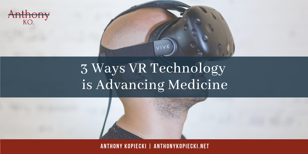
Virtual reality is helping to move the world forward in a number of ways. From cutting down on the space that car dealers need by offering completely VR showrooms, to training drivers and pilots, VR is making the world a better, safer place. VR is also making its way into an industry that may help to save countless lives. VR is showing huge promise in the medical world, both for training, and for surgery as well.
VR in Medical Education
For decades – if not centuries – students have honed their skills on cadavers. The drawbacks are well documented: cadavers are expensive to procure, do not fully simulate work on a living body, and of course, there have always been ethical implications to consider. While VR training should never fully replace training on cadavers, it can significantly decrease the number of human bodies that a school needs to procure. Combining classic cadaver training with VR can allow students to hone their skills while still enabling students to have the experience of handling real human flesh and tissue.
VR in Medical Evaluation
Advanced technology has vastly improved medical imaging in the last decade. So much so, that it can now provide so much detail that it can be time-consuming for physicians to review all of the data that scans can generate. VR technology, however, is capable of assembling the data into a realistic 3D model that can be shared with physicians around the world. A leading heart surgeon in France can view a detailed 3D model of a heart from a patient in China, allowing a multinational team of doctors to make a more accurate diagnosis or understand how best to proceed.
VR in the Operating Room
Contrary to popular belief, every human body has significant variations from every other human body. In the past, surgeons really had no way of knowing what they would find when they cut into a patient. Up until now, scanning has only been helpful in revealing abnormalities in bone and dense tissue. Modern scans are getting better and better at reading soft tissue as well.
With these scans, physicians may be able to create a realistic 3D model of the patient’s internal architecture, which they can then use to train for surgeries. Having a better understanding in advance of precisely what they may expect to encounter when they cut into a patient can help them be better prepared when they do.